Our Published Research
If you would like to make an appointment or have a question, please contact us.
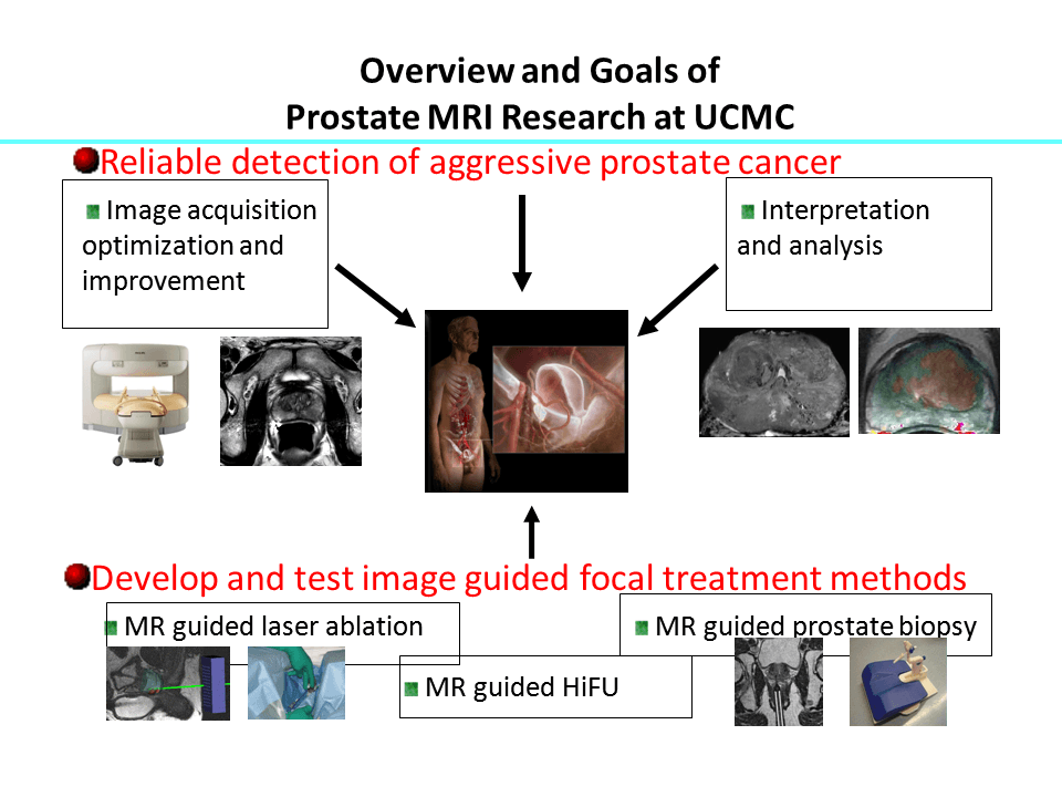
We have a multi-disciplinary research team composed of radiologists, urologists, medical oncologists, radiation oncologists, pathologists, medical physicists and molecular biologists working on developing new imaging and image guided technologies for prostate cancer. The two overarching aims of our research are “non-invasive and accurate diagnosis of aggressive prostate cancer using MR imaging“ and “eradication of localized prostate cancer with minimal complications using minimally invasive treatment methods”. Our group has already has developed new MR sequences, pilot CAD software for prostate MRI and tested MR guided therapy methods such as laser and focused ultrasound ablation in clinical and pre-clinical studies.
Performance of T2 maps in the detection of prostate cancer.
Chatterjee A, Devaraj A, Matthew M, Szasz T, Antic T, Karczmar GS, Oto A.
(In Press- Academic Radiology)
Multi-parametric MRI features and pathologic outcome of wedge shaped lesions on T2-weighted images
Chatterjee A, Tokdemir S, Gallan AJ, Wang S, Yousuf A, Antic T, Karczmar GS, Oto A.
(In Press – American Journal of Roentgenology 18-19742)
Diagnosis of Prostate Cancer with Noninvasive Estimation of Prostate Tissue Composition by Using Hybrid Multidimensional MR Imaging: A Feasibility Study.
Chatterjee A, Bourne R, Wang S, Devaraj A, Gallan AJ, Antic T, Karczmar GS, Oto.
Radiology 2018; 287(3): 864-73.
Evaluation of Focal Laser Ablation of Prostate Cancer Using High Spectral and Spatial Resolution Imaging: A Pilot Study.
Shiyang Wang, PhD, Xiaobing Fan, PhD, Ambereen Yousuf, MD, Scott E. Eggener, MD, Gregory Karczmar, PhD, and Aytekin Oto, MD.
J Magn Reson Imaging. Accepted 2018.
Comparison of T2 weighted, Diffusion Weighted and Dynamic Contrast Enhanced MRI for Calculation of Index Prostate Cancer Volume: Correlation with Whole Mount Pathology.
Sun C, Chatterjee A, Yousuf A, Antic T, Eggener S, Karczmar GS, Oto A.
(In press – American Journal of Roentgenology 18-20147)
Feasibility of Dynamic Contrast-Enhanced Magnetic Resonance Imaging Using Low-Dose Gadolinium: Comparative Performance With Standard Dose in Prostate Cancer Diagnosis.
He D, Chatterjee A, Fan X, Wang S, Eggener S, Yousuf A, Antic T, Oto A, Karczmar GS.
Invest Radiol. 2018 Oct;53(10):609-615.
MRI Findings Following MRI Guided Focal Laser Ablation of Prostate Cancer.
Westin C, Chatterjee A, Ku E, Yousuf A, Wang S, Thomas S, Fan X, Eggener S, Karczmar G, Oto A.
AJR Am J Roentgenol. 2018 Sep;211(3):595-604.
Performance of ultrafast DCE-MRI for diagnosis of prostate cancer.
Chatterjee A, He D, Fan X, Wang S, Szasz T, Yousuf A, Pineda F, Antic T, Mathew M, Karczmar GS, Oto A.
Acad Radiol. 2018 Mar;25(3):349-358.
Comparison of arterial input functions measured from ultra-fast dynamic contrast enhanced MRI and dynamic contrast enhanced computed tomography in prostate cancer patient.
Wang S, Lu Z, Fan X, Medved M, Jiang X, Sammet S, Yousuf A, Pineda F, Oto A, Karczmar GS.
Phys Med Biol. 2018 Jan 30;63(3):03NT01.
MRI evaluation of benign prostatic hyperplasia: Correlation with international prostate symptom score.
Guneyli S, Ward E, Peng Y, Nehal Yousuf A, Trilisky I, Westin C, Antic T, Oto A.
J Magn Reson Imaging. 2017 Mar;45(3):917-925.
Multi-parametric MR imaging of the anterior fibromuscular stroma and its differentiation from prostate cancer
Ward, E., Baad, M., Peng, Y., Yousuf, A., Wang, S., Antic, T., Oto, A.
Abdom Radiol (2016)
Short-term reproducibility of apparent diffusion coefficient estimated from diffusion-weighted MRI of the prostate.
Sadinski M, Medved M, Karademir I, Wang S, Peng Y, Jiang Y, Sammet S, Karczmar G, Oto A.
Abdom Imaging. 2015 Mar 25.
Dynamic Contrast-enhanced MR Imaging Curve-type Analysis: Is It Helpful in the Differentiation of Prostate Cancer from Healthy Peripheral Zone?
Hansford BG, Peng Y, Jiang Y, Vannier MW, Antic T, Thomas S, McCann S, Oto A.
Radiology. 2015 May;275(2):448-57.
Deformable segmentation of 3D MR prostate images via distributed discriminative dictionary and ensemble learning.
Guo Y, Gao Y, Shao Y, Price T, Oto A, Shen D.
Med Phys. 2014 Jul;41(7):072303.
High-resolution diffusion-weighted imaging of the prostate.
Medved M, Soylu-Boy FN, Karademir I, Sethi I, Yousuf A, Karczmar GS, Oto A.
AJR Am J Roentgenol. 2014 Jul;203(1):85-90.
MR imaging of the prostate.
Yacoub JH, Oto A, Miller FH.
Radiol Clin North Am. 2014 Jul;52(4):811-37.
Dynamic contrast-enhanced MR imaging features of the normal central zone of the prostate.
Hansford BG, Karademir I, Peng Y, Jiang Y, Karczmar G, Thomas S, Yousuf A, Antic T, Eggener S,Oto A.
Acad Radiol. 2014 May;21(5):569-77.
Laser ablation as focal therapy for prostate cancer.
Wenger H, Yousuf A, Oto A, Eggener S.
Curr Opin Urol. 2014 May;24(3):236-40.
Apparent diffusion coefficient for prostate cancer imaging: impact of B values.
Peng Y, Jiang Y, Antic T, Sethi I, Schmid-Tannwald C, Eggener S, Oto A.
AJR Am J Roentgenol. 2014 Mar;202(3):W247-53.
Validation of quantitative analysis of multiparametric prostate MR images for prostate cancer detection and aggressiveness assessment: a cross-imager study.
Peng Y, Jiang Y, Antic T, Giger ML, Eggener SE, Oto A.
Radiology. 2014 May;271(2):461-71.
MR imaging of the prostate and adjacent anatomic structures before, during, and after ejaculation: qualitative and quantitative evaluation.
Medved M, Sammet S, Yousuf A, Oto A.
Radiology. 2014 May;271(2):452-60.
Revisiting the central gland anatomy via MRI: does the central gland extend below the level of verumontanum?
Hansford BG, Peng Y, Jiang Y, Al-Ahmadie H, Eggener S, Yousuf A, Oto A.
J Magn Reson Imaging. 2014 Jan;39(1):167-71.
Hybrid multidimensional T(2) and diffusion-weighted MRI for prostate cancer detection.
Wang S, Peng Y, Medved M, Yousuf AN, Ivancevic MK, Karademir I, Jiang Y, Antic T, Sammet S,Oto A, Karczmar GS.
J Magn Reson Imaging. 2014 Apr;39(4):781-8.
MR Imaging of the Prostate and Adjacent Anatomic Structures before, during, and after Ejaculation: Qualitative and Quantitative Evaluation.
Medved M, Sammet S, Yousuf A, Oto A.
Radiology. 2014 February.
Cross-device automated prostate cancer localization with multiparametric MRI.
Artan Y, Oto A, Yetik IS.
IEEE Trans Image Process. 2013 Dec;22(12):5385-94.
Prostate volumes derived from MRI and volume-adjusted serum prostate-specific antigen: correlation with Gleason score of prostate cancer.
Karademir I, Shen D, Peng Y, Liao S, Jiang Y, Yousuf A, Karczmar G, Sammet S, Wang S, Medved M, Antic T, Eggener S, Oto A.
AJR Am J Roentgenol. 2013 Nov;201(5):1041-8.
Revisiting the central gland anatomy via MRI: Does the central gland extend below the level of verumontanum?
Hansford BG, Peng Y, Jiang Y, Al-Ahmadie H, Eggener S, Yousuf A, Oto A.
J Magn Reson Imaging. 2014 Jan;39(1):167-71.
Hybrid multidimensional T(2) and diffusion-weighted MRI for prostate cancer detection.
Wang S, Peng Y, Medved M, Yousuf AN, Ivancevic MK, Karademir I, Jiang Y, Antic T, Sammet S, Oto A, Karczmar GS.
J Magn Reson Imaging. 2013 Aug 1.
Standards of reporting for MRI-targeted biopsy studies (START) of the prostate: recommendations from an International Working Group.
Moore CM, Kasivisvanathan V, Eggener S, Emberton M, Fütterer JJ, Gill IS, Grubb Iii RL, Hadaschik B, Klotz L, Margolis DJ, Marks LS, Melamed J, Oto A, Palmer SL, Pinto P, Puech P, Punwani S, Rosenkrantz AB, Schoots IG, Simon R, Taneja SS, Turkbey B, Ukimura O, van der Meulen J, Villers A, Watanabe Y.
START Consortium. Eur Urol. 2013 Oct;64(4):544-52.
Seminal vesicle invasion in prostate cancer: evaluation by using multiparametric endorectal MR imaging.
Soylu FN, Peng Y, Jiang Y, Wang S, Schmid-Tannwald C, Sethi I, Eggener S, Antic T, Oto A.
Radiology. 2013 Jun;267(3):797-806.
MR imaging-guided focal laser ablation for prostate cancer: phase I trial.
Oto A, Sethi I, Karczmar G, McNichols R, Ivancevic MK, Stadler WM, Watson S, Eggener S.
Radiology. 2013 Jun;267(3):932-40.
Quantitative analysis of multiparametric prostate MR images: differentiation between prostate cancer and normal tissue and correlation with Gleason score–a computer-aided diagnosis development study.
Peng Y, Jiang Y, Yang C, Brown JB, Antic T, Sethi I, Schmid-Tannwald C, Giger ML, Eggener SE, Oto A.
Radiology. 2013 Jun;267(3):787-96.
ACR Appropriateness Criteria prostate cancer–pretreatment detection, staging, and surveillance.
Eberhardt SC, Carter S, Casalino DD, Merrick G, Frank SJ, Gottschalk AR, Leyendecker JR, Nguyen PL, Oto A, Porter C, Remer EM, Rosenthal SA.
J Am Coll Radiol. 2013 Feb;10(2):83-92.
Cross-device automated prostate cancer localization with multiparametric MRI.
Artan Y, Oto A, Yetik IS.
Conf Proc IEEE Eng Med Biol Soc. 2012;2012:6247-50.
Microvessel density is not increased in prostate cancer: digital imaging of routine sections and tissue microarrays.
Tretiakova M, Antic T, Binder D, Kocherginsky M, Liao C, Taxy JB, Oto A.
Hum Pathol. 2013 Apr;44(4):495-502.
Ultrasound- and MR-guided focused ultrasound surgery for prostate cancer.
Zini C, Hipp E, Thomas S, Napoli A, Catalano C, Oto A.
World J Radiol. 2012 Jun 28;4(6):247-52.
Evaluation of the prostate bed for local recurrence after radical prostatectomy using endorectal magnetic resonance imaging.
Liauw SL, Pitroda SP, Eggener SE, Stadler WM, Pelizzari CA, Vannier MW, Oto A.
Int J Radiat Oncol Biol Phys. 2013 Feb 1;85(2):378-84.
Local staging of prostate cancer with MRI.
Soylu FN, Eggener S, Oto A.
Diagn Interv Radiol. 2012 Jul-Aug;18(4):365-73.
Dynamic contrast-enhanced MR imaging findings of bone metastasis in
patients with prostate cancer.
Kayhan A, Yang C, Soylu FN, Lakadamyalı H, Sethi I, Karczmar G, Stadler W, Oto A.
World J Radiol. 2011 Oct 28;3(10):241-5.
Diffusion-weighted and dynamic contrast-enhanced MRI of prostate cancer: correlation of quantitative MR parameters with Gleason score and tumor angiogenesis.
Oto A, Yang C, Kayhan A, Tretiakova M, Antic T, Schmid-Tannwald C, Eggener S, Karczmar GS, Stadler WM.
AJR Am J Roentgenol. 2011 Dec;197(6):1382-90.
High-resolution MRI of excised human prostate specimens acquired with 9.4T in detection and identification of cancers: validation of a technique.
Fan X, Haney CR, Agrawal G, Pelizzari CA, Antic T, Eggener SE, Sethi I, River JN, Zamora M, Karczmar GS, Oto A.
J Magn Reson Imaging. 2011 Oct;34(4):956-61.
Multi-parametric MR imaging of transition zone prostate cancer: Imaging features, detection and staging.
Kayhan A, Fan X, Oommen J, Oto A.
World J Radiol. 2010 May 28;2(5):180-7.
Prostate cancer: differentiation of central gland cancer from benign prostatic hyperplasia by using diffusion-weighted and dynamic contrast-enhanced MR imaging.
Oto A, Kayhan A, Jiang Y, Tretiakova M, Yang C, Antic T, Dahi F, Shalhav AL, Karczmar G, Stadler WM.
Radiology. 2010 Dec;257(3):715-23.
Dynamic contrast-enhanced magnetic resonance imaging in prostate cancer.
Kayhan A, Fan X, Oto A.
Top Magn Reson Imaging. 2009 Apr;20(2):105-12.
Reproducibility assessment of a multiple reference tissue method for quantitative dynamic contrast enhanced-MRI analysis.
Yang C, Karczmar GS, Medved M, Oto A, Zamora M, Stadler WM.
Magn Reson Med. 2009 Apr;61(4):851-9.