Who Needs Prostate MRI Services?
If you would like to make an appointment or have a question, please contact us.

Prostate Cancer: Who is at Risk and
How it is Detected and Diagnosed
Prostate cancer (Pca) is the most frequently diagnosed cancer in males and the second leading cause of cancer-related death in men, causing approximately 40,000 deaths very year. It is more common among African-American men as compared to Caucasian men, and it is usually detected at more advanced stages in African-Americans.
The number of new diagnoses of Pca (~ over 200,000/year) far outweigh the number of lethal cases. Most men with prostate cancer die of other causes, and many tumors – perhaps the majority – would never cause symptoms or result in death if left undetected and untreated.
One in six American men will develop Pca during their lifetime, but one in 34 will die of the disease.
Currently, Pca screening is driven by serum prostate specific antigen (PSA) values and digital rectal examination. The definitive diagnosis is based on histologic analysis of tissue obtained by transrectal ultrasound (TRUS) guided biopsies.
Based on elevated serum PSA results, more than 1.2 million prostate needle biopsies are performed every year in US alone, with an estimated cost to the health care system of 2 billion dollars per year. This places an enormous burden on patients, families, and medical professionals. Despite the high cost and widespread adoption of this screening strategy, the benefit and efficiency of current screening methods is controversial.
What is the Screening Controversy?
The U.S. Preventive Services Task Force recently concluded that PSA-based screening results in small or no reduction in prostate cancer-specific mortality and is associated with harms related to subsequent evaluation and treatments and thus recommended limited use of the PSA test. On the other hand, the American Urologic Association (AUA) recommends that screening be considered for asymptomatic men ages 55 to 69 years. Unfortunately, current diagnostic tools, including PSA screening, are unable to distinguish life-threatening prostate tumors from indolent tumors. As a result, many patients have received unnecessary, invasive prostate biopsies and aggressive therapy, which often has side effects, unnecessary cost, and emotional distress for the patient.
Diagnosing Prostate Cancer
The greatest difficulty for clinicians at the time of diagnosis is in deciding which men have fast-growing cancers that need radical treatment and which have slow-growing cancers that will never trouble them. The most important predictors of prognosis in prostate cancer are Gleason scores (GS) and tumor staging. However, accurate estimation of these parameters is currently only possible following radical prostatectomy.
Biopsy-based Gleason scores are often inaccurate and approximately 30% of tumors have a higher Gleason score on repeat biopsy or in the radical prostatectomy specimen. This is likely due to sampling error, resulting from the inability to identify and target the most significant tumor or tumor region in TRUS guided biopsy.
There is a critical and immediate need for accurate, non-invasive tools to improve diagnosis of prostate cancer, including a non-invasive evaluation of the entire prostate at the time of initial diagnosis to discriminate between high grade disease that requires aggressive treatment and indolent disease. This would have significant benefits for patients, and reduce the financial, logistical, and emotional costs associated with prostate cancer.
MRI of the Prostate Can Improve the Accuracy of the Diagnosis
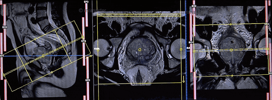
Magnetic resonance imaging (MRI) is currently the best non-invasive tool for imaging cancer in the prostate because of its high soft-tissue contrast, multi-planar capabilities, and has the potential for providing unique functional and biologic information not available with other imaging modalities. Multi-parametric MR imaging, including T2-weighted imaging, Diffusion-weighted MR (DW-MRI) and dynamic contrast enhanced MRI (DCE-MRI) is a widely accepted tool for diagnosis, local staging, and detection of recurrence of prostate cancer (Pca).
MR imaging can potentially be helpful to answer several questions critical for the management of patients with prostate cancer:
- Patients with elevated PSA before biopsy or after initial negative biopsy: MRI can show the location of the cancer (or its lack of) within the prostate in these patients. It has been shown that targeted biopsies using MR information can provide better yield compared to random US guided biopsies. Alternatively, MRI can save the patients from unnecessary biopsies.
- Local staging of Pca for patients who have intermediate risk Pca: MRI can demonstrate extracapsular invasion, seminal vesicle invasion or enlarged lymph nodes. This information can influence the management decisions and allow both the patient and treating physician to make more educated decisions for their management.
- Triage and follow-up patients who chose to undergo Active surveillance (AS): Approximately, one third of the patients who chose to undergo AS may eventually need more radical treatment due to detection of more aggressive disease. MRI can be helpful in early detection of these patients and may have a role in the follow-up patients with AS.
- Treated patients with elevated PSA: Elevated PSA is the initial sign of recurrence after treatment (radiation or prostatectomy). MRI may be helpful in identification of the location (pelvis or metastatic) and extent of the disease and can impact management.
- Focal therapy: Analogous to lumpectomy in breast cancer, focal therapy is an emerging paradigm in treatment of Pca. Accurate localization of the cancer within the prostate using MRI is crucial for a successful focal treatment.